Release date: 2017-10-31
As a technical expert and practical expert in medical imaging, Professor Xing Lei, the director of Stanford University Medical Physics Center and the chief scientist of Huiyi Huiying, has been invited to participate in the domestic and international radiotherapy conferences, including the 2017 American Medical Physics Annual Conference (AAPM). International Symposium on Image Computing and Digital Medicine in Chengdu, the first application of artificial intelligence in medicine, the annual meeting of the American Society of Radiation Oncology (ASTRO), and the branch of the Radiological Specialist of the Beijing Medical Association. AI technology has been widely discussed in recent years. How to integrate AI into clinic, how to help video doctors to release more value, how to use image diagnosis as an entry point, and use AI to penetrate the whole chain of cancer treatment is also a hot topic. This is a game of data and algorithm, but also a diagnosis. Synergistic with the treatment.
Dr. Xing Lei, the head of the Department of Medical Physics at Stanford University and Distinguished Professor of Stanford University, presented the summit of the Human Intelligence Analysis Cloud Platform (English version). The wonderful sharing of Professor Xing's theme "Application of AI in Clinical Diagnosis and Treatment" won the unanimous approval of the majority of participants. Professor Xing Lei is a tenured professor at Stanford University and a professor at Stanford's Department of Electrical Engineering, Molecular Imaging and Bioinformatics, and Bio-X. He has been engaged in medical imaging, medical physics and medical information teaching for more than 20 years. He has published more than 300 professional papers and has hosted major scientific research projects of NIH, DOD, NSF, ACS, RSNA and other institutions. American Cancer Society Research Scholar Award, American Academy of Medical Physics (AAPM) Best Paper Award, and Google Research Award. Dr. Xing Lei is also a Fellow of AAPM and AIMBIE (American Institute of Medical and Biological Engineering) and a national “Thousand Talents Program†expert.
The following is the original collection of Dr. Xing Lei's share. Lei Feng did an editor who did not change the original intention:
Clinical pain points promote the application of AI in medicine
To discuss the application of AI in medicine, we must first understand what medicine is. Modern medicine is Evidence-based Medicine, which consists of three parts: clinical experience, scientific data, and the actual condition and willingness of patients.
These three points are simple to look at, but clinical practice is difficult. Medicine is science and an art, involving the quality and experience of doctors. For this reason, there are many problems in clinical medicine, which create an excellent opportunity for the application of AI in the medical field.
First, clinically collected data often carries certain biases and uncertainties. Using these empirical data to make clinical decisions is a very complicated process. This process is often difficult or impossible to describe with a simple mathematical model. Secondly, the radiology and radiotherapy departments are currently duplicating and labor intensive. Third, the cost of building a medical imaging department is high, and the infrastructure between the major top three hospitals and the municipal and county-level hospitals is very different. And AI technology can expand with the scale of the application, the marginal cost is continuously reduced, and has an unparalleled advantage. The application of AI technology can free doctors from many complicated and inefficient jobs, improve the average level and efficiency of medical workers, and enable them to spend valuable time and energy on more valuable creative clinical work. Further, clinical trials often take a long time, from the results to the actual application of the clinical, often takes three to five years. The application of AI technology can accelerate the efficiency of clinical trials. It is no exaggeration to say that AI is an indispensable technology for individualized medical care.
In evidence-based medicine, clinical decisions are closely linked to evidence and data. With the rapid advancement of science and medical technology, the more data, the more clinically determined dimensions. From the perspective of cognitive ability, the number of variables that a person can consider at the same time is very limited. If you can handle ten factors at the same time, you can count as superman. But in reality, the factors that a cancer doctor should consider often far exceed this dimension, and the difficulty and uncertainty can be imagined.
In addition, we are in an era of knowledge explosion. There are articles published every day, and the half-life of these knowledge is only a few years on average, and it is easy to be forgotten by obsolescence. Therefore, it is very important to apply AI to quickly extract the essence of data for clinical use.
Deep learning in clinical applications
I believe that experts are familiar with computer-aided diagnosis, that is, CAD. As early as the 1980s, there were many people doing this and many companies emerged. R2 Technology, which was later acquired by Hologic Inc., is one of the more famous representatives. With the increased computing power of computers and the emergence of GPUs, deep learning is gradually coming to everyone's attention. Today, deep learning has been widely used in our daily work and life.
The process of machine learning is very similar to the process of child cognition: Train the machine through a large number of existing samples and tell it what the cat is and what the dog is. After learning a certain number of samples, the machine recognizes the kittens and puppies when they see them on other different occasions. There are many scenarios in which this ability is used in real life. A radiologist based on his own experience, making a diagnosis based on the patient's medical history and other clinical information is a classic example.
Machine learning can be divided into three categories. We must first use a large amount of data to train the model before we can analyze and predict the new data. In recent years, deep learning is very hot, which may have a great relationship with the battle of interpersonal Go:). In fact, it is not the first time that a machine has defeated mankind. As early as twenty years ago, IBM's Deep Blue had already defeated Kasparov, the chess king of the chess world. Recently, AlphaGo's two man-machine wars have pushed artificial intelligence to a new level, because Go has always been hailed as "the jewel in the crown of human wisdom."
Deep learning and intensive deep learning are the most widely used technologies in medical imaging. They can be used to solve many problems that were previously impossible to solve. This year's Stanford computer department's S. Thrun's research on skin cancer testing published in Nature can be said to be a very successful case. Based on nearly 130,000 skin cancer samples, they trained a CNN deep learning model and tested it with about 2,000 samples. The performance of this model is comparable to that of experienced dermatologists.
In fully exploiting the potential of artificial intelligence and building a global intelligent medical imaging platform, Huiyi Huiying has always been at the forefront of the industry. Huiyi Huiying is using deep learning to simulate the human brain's understanding of three-dimensional images and has made amazing progress. The human brain processes the image from five dimensions: color, shape, and abstract recognition. Therefore, the algorithms for simulating cognitive processes in different regions are not the same. They have accumulated a great deal of experience in practice and have made excellent progress in matching a large amount of clinical data accumulation, improving computational efficiency, optimizing deep learning algorithms, and building models that are constantly improving themselves.
I. AI predicts side effects of liver cancer lung cancer radiotherapy in treatment plan
AI has many applications in radiotherapy, such as how to predict the possible side effects of liver cancer lung cancer radiotherapy. The deep learning model can be used to replace the existing nomogram to achieve more accurate individualized prediction. The current method is to estimate the toxicity of radiotherapy by some indicators after giving the dose distribution. For example, the average dose is greater than the amount that will produce clinically unacceptable toxicity. Using the deep learning model can replace these old indicators and no longer rely on only a few parameters for clinical decisions.
Using the data of a large number of patients, treatment plans and post-treatment toxicity data, we constructed the world's first deep-learning radiotherapy model based on deep learning. It can easily and accurately predict the prognosis of new patients. Compared with the actual clinically observed prognosis, the prediction of deep learning is much more accurate than the existing model. This can be said to be the first substantive application of deep learning in radiotherapy transformation medicine.
Second, the application of AI in treatment planning and image analysis reconstruction
The radiotherapy process is very individual and needs to be continuously optimized based on the patient's anatomy and tumor location. This is a very complicated process. Therefore, developing a treatment plan is a very time consuming task. In general, it takes a few hours to a few days for an experienced technician to develop a treatment plan for a complex patient. Our department has about 3,000 patients each year, and we can imagine how much time and effort it takes. Using deep learning to develop a treatment plan can greatly improve the efficiency and quality of the treatment plan. Currently, we have successfully applied Google AlphaGo's algorithm to the optimization of treatment plans. We compare the treatment plan developed by this algorithm with the manual plan, and the result is better than the plan generated by the existing method, and it is easy to implement on the accelerator.
MRI is used to image moving organs such as the heart or to use MRI-guided radiotherapy, and MRI images need to be acquired and reconstructed in real time in real time. Existing MRIs can generate 4-8 frame images per second. The deep learning model can greatly shorten the time required for 3D MRI image reconstruction, making "real-time" 4D MRI image reconstruction possible. Machine learning can also be applied to the reconstruction of CT images. In CT imaging, the patient usually receives a radiation dose of 1-5 cGy. If the dose is reduced, the noise signal will increase significantly, resulting in a decrease in image quality. We used a previous patient's CT data to construct a model that was used in conjunction with new low-dose data for reconstruction. Thereby the radiation dose of the CT imaging will be greatly reduced.
Third, the application of AI and influence group in image analysis and clinical application
The application research of deep learning in disease screening and detection is also very active, and new methods and new technologies are emerging one after another. For example, our lab is using deep learning to improve existing prostate cancer detection methods. Roughly speaking, when prostate-specific antigen (PSA) is found to be elevated, MRI and biopsy are generally used to confirm the diagnosis. Through a large number of patient images and diagnostic results, we use deep learning to conduct a diagnostic analysis of prostate tumors, thereby finding out the extent of deterioration of all lesions and tumors, thereby avoiding or reducing the cost and pain of biopsy and patients.
I believe that everyone is familiar with radiology. Speaking of radiology, I will introduce a book to you, entitled "Radiomics and Radiogenomics", which was written by my three colleagues at Stanford and will be published early next summer. Using radionomics, eigenvalues ​​can be screened. We recently published an article on radiology to study the prognosis of gliomas in Radiology. In addition, a very important clinical problem in the treatment of glioma is how to distinguish between false progress and true progress. In the regular review of patients after treatment, the treatment decisions for the above two conditions are completely different - the former should be discontinued and the latter should continue treatment. Using neural networks should be a good solution to this problem.
Challenges encountered by AI in clinical applications - algorithms, data, and data exchange methods
So far, the research applications of computer vision have been mainly carried out in two-dimensional space. Medical images are almost all three-dimensional or even four-dimensional, such as CT, MRI, PET and so on. The real medical image learning and processing is just getting started. In addition to algorithms, another key to the application of AI in the medical field is data and how to efficiently exchange data. Deep learning requires constant improvement of the model and therefore requires massive amounts of data. The Huihui Huiying platform can store data in the cloud in a format that everyone agrees for, and is shared by multiple experts - this is crucial for big data processing and deep learning. In addition to image and electronic medical record (EMR) data, we hope to integrate patient genomics data and wearable data into the future, making it easier to conduct multi-faceted deep learning. Data sharing is actually a big problem, involving technology, management, and society. An article published in Lancet Oncology has been discussed in depth, and the barriers to data sharing mentioned in it are also common in China. I believe that with the large amount of high-quality data coming out, the future of AI in clinical applications is very optimistic.
Finally, summed up, today I mainly discuss the application of deep learning in diagnosis, image reconstruction, radiotherapy decision-making. At present, the application of artificial intelligence in the medical field has just begun, and it takes three to five years to truly realize the substantive application of clinical medicine. There is still a long way to go in the future. As for whether AI can replace the "eternal topic" of doctors in the future, we will discuss it in a more relaxed environment later :)
Source: AI Nuggets (Micro Signal HealthAI)
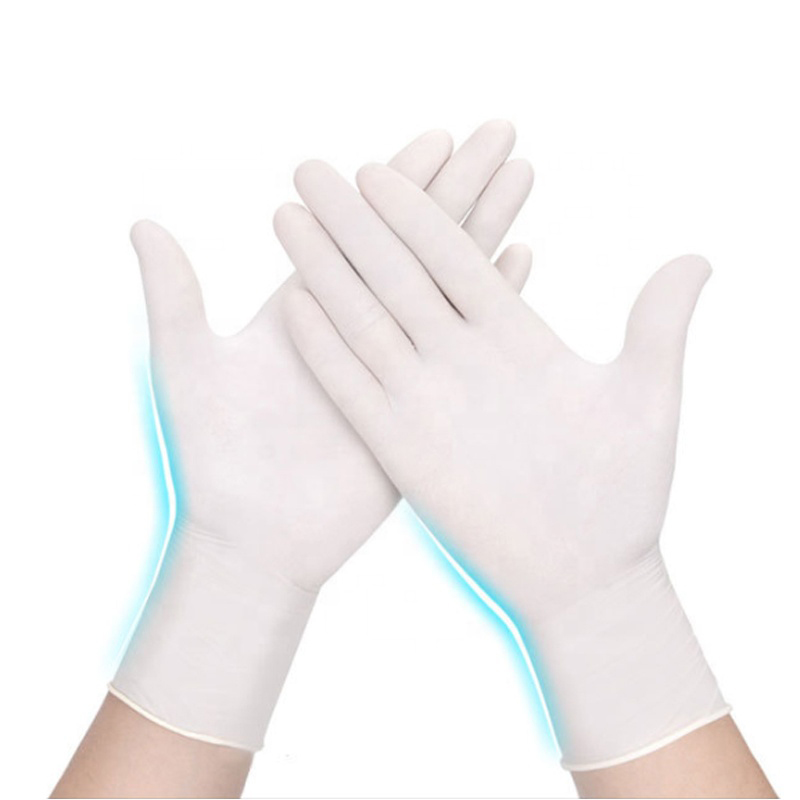
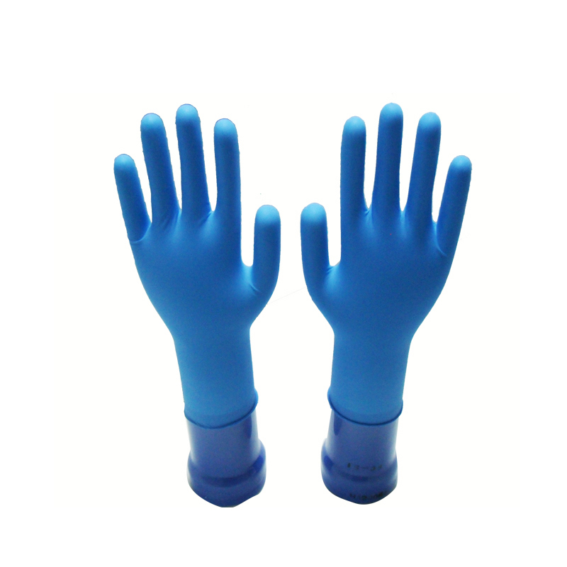
Viruses and bacteria are everywhere. Preventing them in advance is the best way to reduce the risk of infection. Wearing a mask and washing hands frequently are crucial. The new type of coronavirus has the characteristics of contact transmission, and the fact that the virus exists on the doorknob of patients with pneumonia has also shown the necessity of paying attention to hand hygiene and wearing disposable protective gloves.
Whether it is going out to purchase daily necessities or buying through online platforms, it is inevitable to come into contact with "foreign" items, such as supermarket shopping carts, packaging bags, express packaging boxes, etc., and more things need to be touched after resuming work, such as It is inevitable to take public transportation, elevators, etc., these places may have been touched by patients or suspected patients. Wearing disposable protective gloves is an effective means to reduce the risk of infection when there is no condition to wash your hands at any time.


Medical Glove,medical nitrile gloves,Disposable examination medical gloves,surgical gloves
Shanghai Rocatti Biotechnology Co.,Ltd , https://www.shljdmedical.com